Sunday, June 7, 2015

Lipoprotein(a) and macrophages in tandem within a human coronary plaque biopsy:

Here are some pictures of Lp(a) together with macrophages taken from a human coronary atherosclerotic plaque.  Panel C are the macrophage foam cells, Panel D is Lp(a).  They are clearly one on top of the other.
This is taken from: Lipoprotein(a) and inflammation in human coronary atheroma: association with the severity of clinical presentation  from all the way back in 1998.  It would be inaccurate to say that there is only 1 person on the planet who recognizes Lp(a) as an atherogenic factor.

The question of causality and time-frame is always to question.  The essential point we're trying to make is that Lp(a) doesn't just swoop in from out of nowhere for no reason all the sudden.  Macrophages which do home into inflammatory cytokine signals emitted by the artery, and do burrow in to receive cholesterol via active receptor exchange to return to the liver, also don't just suddenly go in and start chewing up lipoproteins.  That being said, foam cells and Lp(a) happen together frequently.  In any remodeling process in the body, there is nearly certainly found macrophages.  You can't just ignore the immune system.

What is my position?  It is irrelevant.  It is beyond irrelevant "who is right," but what is germaine is "WHAT is right."  I do not care "who" is right, nor should you.  Those who care intensely about who should be right obviously have something else driving them like a jerk's ego or some frivolous vendetta.  The question should be, how do we make each and every one of the millions of people of Earth right in their application of science.  What we should all care about is "what is right," not "who is right."  Needless to say, that is my concern in science, no extraneous nonsense.

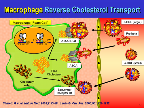
What is true is that macrophages are initially participating in a beneficial remodeling process, and not chewing up ox-LDL in some parasitic process, but attempting to hand back the remodeling waste, the construction site debris back to the liver for recycling via....HDL.  We certainly can't ignore HDL in this repair process either.  It will spin cardiopathology's noodle when they see that the majority of atherosclerotic plaques are rich in ApoE, which if extended the old logic applied to LDL is causing harmful things.  On the contrary, the presence of ApoE we all know to be part of the repair process which may be overwhelmed by a net excess of damage as opposed to a net excess of repair.

Does Lp(a) cause atherosclerosis?  Only if there is a reason for atherosclerosis to take place.  For a many humans who eat just an orange a day but still have the Gulo-/- genetic defect, there is a lot of cumulative damage that does occur which needs patching by Lp(a) and the coagulation system.  Think of it this way, when thinking about preserving arterial integrity: a normal rat makes the equivalent of 5 grams a day in a 70 kilo human when nothing is going on.  In stressful times, a rat makes 300% more vitamin C.  This entry certainly is not encouraging or supporting the fallacy that dietary cholesterol causes heart disease.  It does not, but other dietary factors such as too low vitamin C, B, E, magnesium, K, D, amino acids and too much trans-fatty acids and omega-6 do.  The recent US Federal guidance to stop paying attention to ingested cholesterol is a well-studied one made with decades of clinical evidence that cholesterol consumption has historically had nothing to do with the rate of heart disease.

I think this statement I made a long time ago sums it up nicely about what is going on:

 "A big lump is better than a big hole."

One prevents lethal hemorrhage, the most dramatic example in human biology being the aortic dissection with adventitial failure, the other is lethal hemorrhage.  One of the first priorities of the human physiology is to stop bleeding and hemorrhaging.  It drops everything it is doing and attends to that first, even if it means making a pile of disorganized stuff at the site of leaking that is harmful down the road.  Better than bleeding to death.  Lp(a) is very atherogenic at sites of arterial damage, and sticks more to the glycocalyx with ascending concentrations.  If the endothelium is in tact, there is nothing for Lp(a) to react to, no ligand, no binding site to the ligand.  Lp(a) just floats along harmlessly as it does not encounter plasminogen binding sites, free lysyls, fibrin, exposed subendothelial fibronectin, etc.  You can't just ignore the coagulation system either.  Lp(a) has a direct affinity to fibrin(ogen) which is Clotting Factor 1, and gets cross-linked to fibrin clots via FactorXIII.  Do macrophages foam cells CAUSE atherosclerosis?  No.  They are attempting to prevent it, but the process can and does go wrong if they get overwhelmed.  When they are participating in arterial repair, foam cells are formally defined as early plaques, but that is simply a matter of scientific semantics.  Perhaps one day, they will be relabeled as "reparative cells" distinct from a fibrofatty mass devoid of cells that can rupture and cause thrombosis. Given an equal sized foam cell lesion and fibrofatty mass with thin cap that is  less than 20% occlusive, the unstable fibrofatty mass is the dangerous thing, and the foam cell lesion is unconcerning.

Macrophages and foam cells are not always in human atherosclerotic plaques.  In fact, many times, they are not there at all.  Many pathologist specimens of human diseased atherosclerotic arteries do not have foam cells, which is why some scientists choose to de-emphasize them or diminish their importance.  But they do happen, and we can't just ignore them.  One improvement in the future state-of-the-art may be to put macrophages and foam cell "lesions" in a category all on their own as they are dynamically different from all other component atherosclerotic plaques, and not just different in cellular origin and composition.  They also serve an entirely different purpose altogether than other cells found in human plaques, including remodeling, debris clearance, and returning the artery back to its original state.


Saturday, June 6, 2015

You can't just ignore foam cells. Fatty streaks happen, with or without Lp(a). An addendum of "Common Sense."

Given that the guinea pig DOES NOT have Lp(a) but only LDL in a higher ratio to HDL than in wild mice, and that they do incur atherosclerosis upon chronic scurvy, there is no ambiguity that vitamin C deficiency is causal to atherosclerosis WITHOUT invoking apo(a).  Unless a genetically modified variant of guinea pig happens, they will never express apo(a) protein.  The "immunological evidence" proferred by others is obviously a cross-reaction to a different protein, perhaps even apo(a)-like, but certainly it is not lipoprotein(a).  It could not be, rendering any reference to Lp(a) an illogical train of thinking.  However, they do have an ample amount of LDL.  ApoB particles are important to atherogenesis.  We can't just suddenly start ignoring ApoB, not the Lp(a) platform people either.  Without ApoB synthesis, amplified by inflammatory arterial cytokines, there is NO Lp(a).  In mice that can't make vitamin C but also don't have much LDL but a vast majority of HDL, atherosclerosis is limited to a mild physiological hypertrophy, proteoglycan accumulation, and fibrosis, called arteriosclerosis in olden days.  There are some thoracic aortic lesions especially at bifurcations to the digestive system, but little to no atheroma of any visible or clinically significant nature with a majority HDL and few ApoB particles/absolute mass/LDL-cholesterol.  This clearly shows that ApoB containing particles outnumbering ApoA containing particles is a precondition to scurvy induced atherosclerosis, as much as scurvy is a precondition to endothelial dysfunction, the cause and initiation of most atherosclerosis.  This is without getting into the complicated vasa vasorum component which later contributes to erythrocyte evasation into pre-existing plaques where they die and rupture, releasing membrane cholesterol and iron. (It may be in the very near future, the lipid cores and free cholesterol are found to be from intra-plaque hemorrhage from the interdigitating vasa vasorum rather than the vexing (to some people) foam cell plaque "bust.")

That being said, the atherosclerosis that occurs without Lp(a) is much milder than that which occurs with Lp(a).  Atherosclerosis with low Lp(a) and human-esque levels of LDL is much decreased in lesion number, lesion size, and lesion complexity compared to atherosclerosis that occurs with even moderate Lp(a) around.  Lp(a) over 30mg/dL certainly will drive more atherosclerosis, but lower levels of Lp(a) below 20mg/dL also contribute by virtue of its "homing missile" like quality of binding to areas of damage which it finds in the system circulation of 60,000 miles.  It may not matter how much over a certain threshold of Lp(a), but it does potentiate worse and more with ascending blood levels of Lp(a).  There is no question that Lp(a) is atherogenic, but without LDL, how can there be Lp(a)?  It is not an "either-or" or "better or worse" proposition, but where all these facts that the 1000's of highly intelligent global cardiology scientists have seen intersect and what that nexus and conjunction of truth is.

Ox-LDL is a reliable marker of vessel disease and number of vessel involvement

Does this mean that Ox-LDL causes atherosclerosis?  Yes AND No.  It is too low to ever cause atherosclerosis, with circulating levels in multi-vessel CAD being at most 4mg/dL, and normally being 1mg/dL.  However, it is an extremely reliable indicator of CAD, with a level of 3mg/dL Ox-LDL most certainly revealing plaque in the coronaries somewhere.  Its importance is locally in microenvironments where inflammation causes ROS which causes oxidation and acetylation of LDL in the microscopic milieu around the lesion.  If LDL is not oxidized or denatured, it is not "irritating" to the artery, no.  Regular LDL, even in very high concentrations has been proven not to be cytotoxic to the endothelial cell or cause stress fiber contraction.  As I have already said a long time ago, the LDL gets into the artery if and when the endothelial layer (or vasa vasorum) is compromised and has spatially large enough breaches for it to go through.  Vesicular transport would cause LDL to accumulate inside a cell, not outside it, which we'll get to in just a second.

LDL consumption by a cell is highly regulated, and a cell stops internalizing LDL when it has enough, or the cell will turn down its internal synthesis while maintaining internalization.  Extracellular aggregates of native LDL could not be there if they could not reach these subendothelial compartments somehow (increased vascular permeability).  Ox-and Ac-LDL has a special receptor, scavenger receptor, evolutionarily built in humans, the creature with nearly exclusively the majority of fatal heart attacks and strokes, to get rid of apoptotic cells...and denatured LDL.  When scavenger receptor cells encounter ox-LDL, they engulf it without stop.  Call it gobbling, but more accurately, they are the "trash men," the "recycling team," not "police men."  As much as a policeman can clear debris on a highway, and often does for the public benefit, the macrophage can also serve to clean the artery, not "eat it" or "police" it.  The "old concept" people know that fatty streaks, cellular foam cell masses DO NOT burst.  So, the cartoon does call out an inaccuracy - the notion that foam cell lesions form, burst open, and evulse their cholesterol into the artery.  Early on, no.  These are most stable cellular accumulations which would prevent further destabilization of an artery.  Later on, if the condition is unresolved, the damage unmended, and the "artery tumor" becomes hypoxic, like in a tumor, the macrophages would undergo necrosis and form a necrotic core, just like happens in a tumor core.

In human plaques, foam cell formation from the smooth muscle cell component is vastly underestimated because in mice, the majority of foam cells occur from macrophages.  In humans a greater proportion of foam cells happen from the artery cells themselves.  Yet, make no mistake, foam cell "fatty streaks" are found all the time in human atherosclerosis.  They may be there, they may not be, but an astute scientist does not simply ignore foam cells, especially those colocalized upon an Lp(a) deposit.  There are vastly more human plaques that do not have a foam cell component, and these actually are more worrisome because they are less revertible than a foam cell fatty streak.  Why?

The foam cell, like the apo(a) protein, isn't just some random superfluous curiosity, an accident of biology with no purpose.  Foam cells not only contribute to the artery structure temporarily as additional cells with their hydrophobic seal against hemorrhage, cholesterol, but also are participating in transport of excess unused or remaining cholesterol back to the liver, just as LDL delivers useful cholesterol for cell replacement.  Of all cell types, the macrophage is very efficient at this process.  So we see that this too is a useful, purposeful event, that if resolves satisfactorily, signals the macrophages to exit the artery wall, making the fatty streak lump vanish.  Some call it "reverse cholesterol transport," others don't like that name and call it something else.  Once again, you can't just ignore foam cells or dismiss fatty streaks.  Just as Goldstein and Brown didn't just fabricate a mythic fable of LDL homeostasis, despite this encouraging systemic statin poisoning, scientists and pathologists did not 'just make up' a fable of fatty streaks and foam cells being in human arteries.  Truly, these are about repair, rather than a nonsensical auto-attack on the artery wall, although auto-immune arteritis is a real disease too.  It is increasingly important to stay updated with the field to see others' understanding of the issue so that all science and scientists everywhere can eventually come to a real-world consensus.  Or we can rage around ham-fisted and grandstanding, pretending to know everything without the diligence necessary to know everything.

Macrophage reverse cholesterol transport: key to the regression of atherosclerosis?


Sunday, February 1, 2015

Collagen IV derived "tumstatin" suppresses tumor growth.

Collagen IV, the basement membrane collagen is probably one of the oldest and most examined subtypes of collagen in oncology.  It is certainly not the only or most important collagen in cancer suppression, or other diseases, but Col4 has been proven to function as a barrier against metastasis in the fibrous capsule in several sophisticated and technically up-to-date studies.

Inhibition of tumor angiogenesis by tumstatin: insights into signaling mechanisms and implications in cancer regression.

"Sudhakar A1, Boosani CS.

Growing tumors develop additional new blood vessels to meet the demand for adequate nutrients and oxygen, a process called angiogenesis. Cancer is a highly complex disease promoted by excess angiogenesis; interfering with this process poses for an attractive approach for controlling tumor growth. This hypothesis led to the identification of endogenous angiogenesis inhibitors generated from type IV collagen, a major component of vascular basement membrane (VBM). Type IV collagen and the angiogenesis inhibitors derived from it are involved in complex roles, than just the molecular construction of basement membranes. Protease degradation of collagens in VBM occurs in various physiological and pathological conditions and produces several peptides. Some of these peptides are occupied in the regulation of functions conflicting from those of their original integral molecules. Tumstatin (alpha3(IV)NC1), a proteolytic C-terminal non-collagenous (NC1) domain from type IV collagen alpha3 chain has been highlighted recently because of its potential role in anti-angiogenesis, however its biological actions are not limited to these processes. alpha3(IV)NC1 inhibits proliferation by promoting endothelial cell apoptosis and suppresses diverse tumor angiogenesis, thus making it a potential candidate for future cancer therapy. The present review surveys the physiological functions of type IV collagen and discovery of alpha3(IV)NC1 as an antiangiogenic protein with a comprehensive overview of the knowledge gained by us towards understanding its signaling mechanisms."

Tumstatin, the NC1 domain of alpha3 chain of type IV collagen, is an endogenous inhibitor of pathological angiogenesis and suppresses tumor growth. 

"
Angiogenesis, the formation of new blood vessels, is required for physiological development of vertebrates and repair of damaged tissue, but in the pathological setting contributes to progression of cancer. During tumor growth, angiogenesis is supported by up-regulation of angiogenic stimulators (pro-angiogenic) and down-regulation of angiogenic inhibitors (anti-angiogenic). The switch to the angiogenic phenotype (angiogenic switch) allows the tumors to grow and facilitate metastasis. The bioactive NC1 domain of type IV collagen alpha3 chain, called tumstatin, imparts anti-tumor activity by inducing apoptosis of proliferating endothelial cells. Tumstatin binds to alphaVbeta3 integrin via a mechanism independent of the RGD-sequence recognition and inhibits cap-dependent protein synthesis in the proliferating endothelial cells. The physiological level of tumstatin is controlled by matrix metalloproteinase-9, which most effectively cleaves it from the basement membrane and its physiological concentration in the circulation keeps pathological angiogenesis and tumor growth in check. These findings suggest that tumstatin functions as an endogenous inhibitor of pathological angiogenesis and functions as a novel suppressor of proliferating endothelial cells and growth of tumors."


Collagen XIX is strongly anti-tumorigenic

The NC1 domain of type XIX collagen inhibits in vivo melanoma growth.

Mol Cancer Ther. 2007 Feb;6(2):506-14.
The NC1 domain of type XIX collagen inhibits in vivo melanoma growth.
Type XIX collagen is a minor collagen that localizes to basement membrane zones, together with types IV, XV, and XVIII collagens. Because several NC1 COOH-terminal domains of other chains from basement membrane collagens were reported to exhibit antitumor activity, we decided to study the effects of the NC1(XIX) collagen domain on tumor progression using an experimental in vivo model of mouse melanoma. We observed a 70% reduction in tumor volume in NC1(XIX)-treated mice compared with the corresponding controls. Histologic examination of the tumors showed a strong decrease in tumor vascularization in treated mice. In vitro, NC1(XIX) inhibited the migrating capacity of tumor cells and their capacity to invade Matrigel. It also inhibited the capacity of human microvascular endothelial cells to form pseudotubes in Matrigel. This effect was accompanied by a strong inhibition of membrane type-1 matrix metalloproteinase (matrix metalloproteinase-14) and vascular endothelial growth factor expression. Collectively, our data indicate that the NC1 domain of type XIX collagen exerts antitumor activity. This effect is mediated by a strong inhibition of the invasive capacities of tumor cells and antiangiogenic effects. NC1(XIX) should now be considered as a new member of the basement membrane collagen-derived matrikine family with antitumor and antiangiogenic activity.
PMID: 17308049
 

The NC1 domain of type XIX collagen inhibits melanoma cell migration. 

Eur J Dermatol. 2010 Nov-Dec;20(6):712-8. doi: 10.1684/ejd.2010.1070. Epub 2010 Sep 14.
 The NC1 domain of type XIX collagen inhibits melanoma cell migration.
Type XIX collagen is a minor collagen that localizes to basement membrane zones. We previously demonstrated that the C-terminal NC1 domain of type XIX collagen inhibits tumor growth in vivo. In the present study, we analyzed the effects of the NC1(XIX) collagen domain on migratory behaviour of melanoma B16F10 cells. We found that NC1(XIX) do not inhibit melanoma cell proliferation. On the contrary, NC1(XIX) strongly inhibited the migratory capacities of melanoma cells in the scratch wound model and in Ibidi® devices: cell migration speed was 7.69 ± 1.49 μm/h for the controls vs 6.64 ± 0.82 μm/h for cells incubated with 30 μmol/L NC1(XIX) and 5.72 ± 0.67 μmol/h with 60 μmol/L NC1(XIX). Similar results were obtained with UACC 903 human melanoma cells. Further work will be necessary to elucidate the molecular mechanisms of this migration inhibition. It may, however, explain, at least partially, the inhibition of tumor growth that we observed in vivo.
PMID: 20840910


Loss of Collagen XV Causes Muscle Tissue and Capillary Degeneration

Lack of type XV collagen causes a skeletal myopathy and cardiovascular defects in mice.

From:
Proc Natl Acad Sci U S A. 2001 Jan 30;98(3):1194-9. Epub 2001 Jan 23.

Lack of type XV collagen causes a skeletal myopathy and cardiovascular defects in mice.


Muscle histology: focal areas of degeneration, regeneration, and variation in fiber size. Hematoxylin and eosin-stained sections of the gastrocnemius (A), triceps brachii (B), paraspinal (C), and quadriceps (D) muscles of 6-month-old null mice showing cell degeneration (curved arrows, A and C) with macrophage infiltration (A), regenerative fibers (thin arrow, B), central nuclei (thin arrow, C), and increased variation in fiber size with atrophic muscle fibers (D, arrowheads) compared with age-matched wild-type mice (E). Original magnifications: A and D, ×300; B, ×200; C and E, ×80.


Ultrastructural changes in capillaries. Electron microscopy of heart capillaries from a wild-type (A) and a null mouse (B) with swollen endothelial cells (asterisks). The lumen is indicated by a white arrow. Skeletal muscle capillaries from a wild-type (C) and a mutant mouse (D) showing luminal narrowing and folding of endothelial cells.


Effect of isoproterenol stimulation on developed pressure in (A) 6-month-old and (B) 12-month-old mutant and wild-type mice. The responses to β-agonists were measured in terms of the ratio of the maximal value of the developed pressure (DP) to the basal level before the isoproterenol perfusion in Col15a1−/− (●: A, n = 7; B, n = 11) and Col15a1+/+ (□: A, n = 7; B, n = 8) mice. The symbols represent mean ± SEM. Significant differences between the Col15a1−/− and Col15a1+/+ mice are indicated by asterisks as follows: *, P < 0.05, **, P < 0.01. The basal pressure was 31.7 ± 5.0 and 27.3 ± 5.0 mmHg for the 6-month-old control and null mice, respectively, and 20.2 ± 3.4 and 20.8 ± 2.0 mmHg for the 1-year-old mice (mean ± SEM).


Sunday, January 25, 2015

Partial and Total Loss of Arterial Collagen XVIII Causes Atherosclerosis

Loss of Collagen XVIII Enhances Neovascularization and Vascular Permeability in Atherosclerosis

Some factions claim that loss of arterial collagen is causal to atherosclerosis without being very specific about the mechanism (where? the adventitia? the endothelial basement?  The media?  how? immune side?  smc autophagic?  oxidative?  proteolytic? autolysis?) and refer to studies done before regarding deranged hepatic metabolism of cholesterol during scurvy which causes a certain hyperlipidemia by bile metabolism deficiency and consequent lack of fecosteroid excretion.  Others will look at ratios of Collagens I, III, IV, and even V in the atherosclerotic plaque and their ratios during atherosclerosis.  However, the specific mechanism of collagen loss and atherosclerosis has never been scientifically described in a definitive study until this one.  Through simple logic, (common sense), it follows that singular or combinatory losses of the other arterial collagen subtypes would provide the same result experimentally, either in vitro or in vivo.

The Maeda paper showing that scurvy causes Evans Blue extravasation and gross damage to the arterial wall did not describe any atheromas, hyperplasias, or plaques because the mice die from scurvy before they can incur cumulative atherosclerosis.  They did prove that scurvy causes arterial degradation to the extent that lipoproteins and anything else can enter the artery that can't transgress a healthy, vitamin C (ascorbate) replete artery wall.

The implied connection between atherosclerosis and scurvy is collagen.  While ascorbate isn't the only vitamin involved in collagen synthesis, it is the only one that when absent will prevent proper polymerization of hydroxyl residues.  There is nothing to debate in this regard.  No ascorbate = no assembled collagen.  There may be pro-collagen formation and its strands, but they will never be assembled correctly into their "stronger than steel" fibers without vitamin C, but remain a useless stockpile of components.

It is important to note that the correct ratios of the various collagens is exceedingly important.  While much focus has been made on COL4 basement membranes in conferring tensile strength and resistance to metastasis, too much of COL4 without the other collagens in balance can cause mitochondrial death by integrin signaling abnormalities, as well as aberrant vitamin A processing.  It is not as simple as to say "the more collagen the better."  Not whatsoever is it that simple, and such a conclusion could be disastrous in clinic.  Fibrosis is a disorder of too much of the wrong collagen in the wrong place at the wrong time and is a devastating disorder. 

However, if the intrinsic collagens in the artery, such as COLXVIII, vanish, arteries become much more prone to lipid intrusion and the chain of events that occur after that.  As such, the normal constituency of arterial collagens, many of them not even recognized by conventional histopathology stains and immunohistochemistry, prevent large particle lipoprotein intrusion.  Collagen XVIII is a subtype that is virtually ignored in cardiology, yet possibly one of the most important subtypes.  It logically follows that deficiencies in the most commonly acknowledged collagens of the arterial media and basement membrane, COLI, COLIII, and COLIV, would cause similar problems of pathological permeability to both the clotting cascade biomolecules as well as the whole gamut of lipoproteins.  This deposition of foreign matter is understood for over five decades as being the causal moment of atherosclerosis, but it is still not widely accepted in 2015 that the collagenous and protein barrier of the artery built by vitamin C dependent enzymes is important to prevent this permeability event.


Friday, December 19, 2014

Cholesterol in the CNS: Right Place At The Right Time

Cholesterol is indispensable to the brain and nerves.  Native non-oxidized cholesterol is much more important to the nervous system than other organ systems for which such normal levels of cholesterol may be excessive in that system.  So much so that the brain has its own isolated supply separated from the blood by the notoriously hard to cross blood brain barrier (BBB) which relies on collagen to make it selectively impenetrable to certain molecules.  Any excess is turned into a hydroxy- soluble form which is released from the brain into the blood circulation.  However, when it runs low for whatever reason, cholesterol can be and is actively transported across the BBB from the hepatic supply.  Nowdays, the statin industry has introduced some confusion by entering into the science of Alzheimer's disease.  The big million dollar question is how interfering with the mevalonate pathway can help reduce the causal protein tangles when lowering cholesterol below normal is very toxic to neural function.  There are some elucidations regarding isoprenoids involvement, independent of cholesterol.  Perhaps in some instances with specific ApoE isoforms this is true, but in the majority of instances lowering CNS cholesterol damages it instead of benefitting it.  The tangible outcome of cholesterol depletion in the human brain are problems like amnesia, and the very symptoms of Alzheimer's itself.  The question to answer is probably what optimal cholesterol synthesis is to the brain, as compared to that in the blood supply, just as the "normal" brain ascorbate pool (2000-10,000uM)  is much much higher than that of the bloodstream (60-150uM).

Brain-Derived Neurotrophic Factor Regulates Cholesterol Metabolism for Synapse Development

Vitamin C function in the brain: vital role of the ascorbate transporter SVCT2